
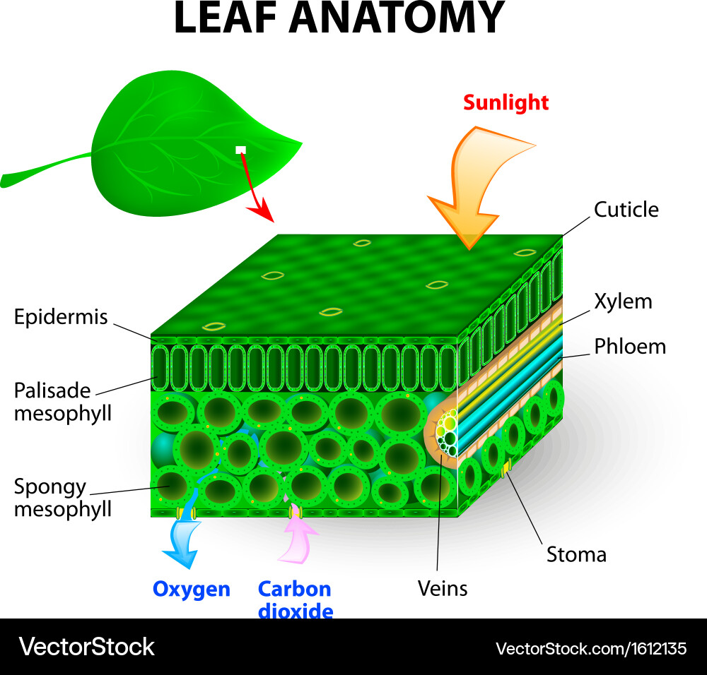
Leaf anatomy vector illustration diagram Anatomy, Medical illustration, Plant science
Whorled Leaf Arrangement; In angiosperm anatomy, a leaf can be identified by where it emerges from the node. In a node, a leaf emerges below the axillary bud. Figure \(\PageIndex{1}\): A diagram of a node. Each node is comprised of a leaf (on the bottom) and an axillary bud (on the top, located in the axil where the petiole meets the stem).
Leaf Structure & Evolution Digital Atlas of Ancient Life
Introduction Leaves are part of the shoot system of the vascular plant sporophyte and one of the three major vegetative (non-reproductive) organs types found in vascular plants (the others are stems and roots). The primary function of leaves is to carry out photosynthesis. Photosynthesis is the process by which a plant makes its food.

Ts Of Dicot Leaf Diagram Amyhj
Figure 9.3. 2: Cross section of a hydrophytic leaf. Observe a prepared slide of a hydrophyte, such as Nymphaea, commonly called a water lily. Note the thin epidermal layer and the absence of stomata in the lower epidermis. In the spongy mesophyll, there are large pockets where air can be trapped.

Labeled Diagram Of A Leaf hubpages
Figure 30.8.1 30.8. 1: Parts of a leaf: A leaf may seem simple in appearance, but it is a highly-efficient structure. Petioles, stipules, veins, and a midrib are all essential structures of a leaf. Within each leaf, the vascular tissue forms veins. The arrangement of veins in a leaf is called the venation pattern.

gleeson11biology / C6
The anatomy of an umbrella tree leaf, of the entire transverse section, with major tissues identified, and a detail of palisade parenchyma cells Umbrella Tree Palisade Cell and Stomata Image on Left - Below: Transmission electron microscope photograph of the palisade parenchyma cell, showing chloroplasts with dark grana stacks and the large.
Leaf Anatomy
[Figure1] Epidermis covers the upper and lower surfaces of the leaf. Usually a single layer of tightly-packed cells, the epidermis mediates exchanges between the plant and its environment, limiting water loss, controlling gas exchange, transmitting sunlight for photosynthesis, and discouraging herbivores.

Parts Of A Leaf Worksheet Printable Worksheets and Activities for Teachers, Parents, Tutors
Book: General Biology (Boundless) 30: Plant Form and Physiology

how to draw a leaf and label it Golden Memoir Photo Gallery
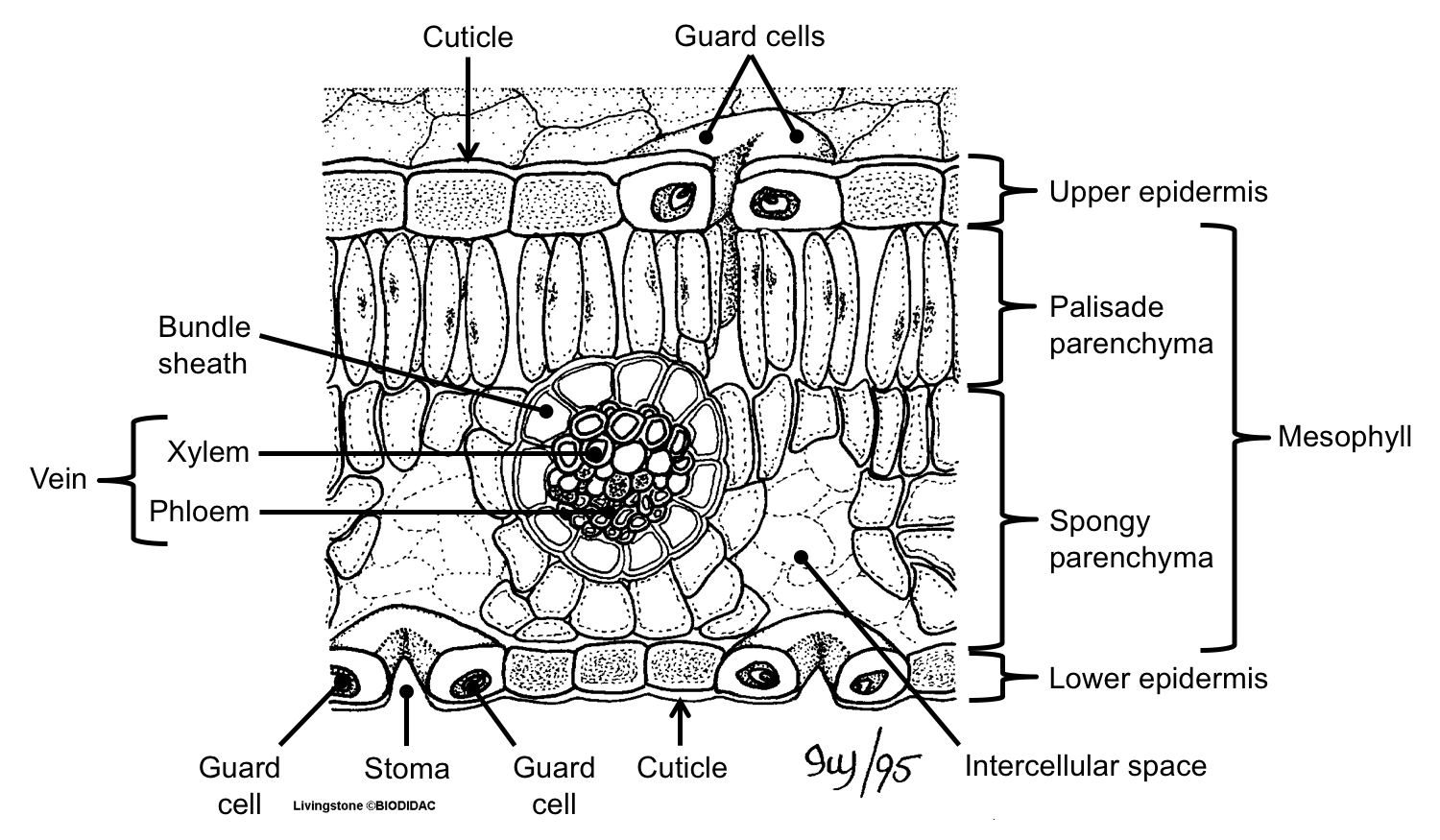
A typical dorsiventral leaf e.g., mango, in transverse section, shows the following structures: [A] Epidermis: It is formed of a single layer of cells, which are closely fitted and have outer thick walls. The outer wall is usually cutinized. The chloroplast and stomata generally not present. [B] Mesophyll:
:max_bytes(150000):strip_icc()/parts_of_a_leaf-56abaed23df78cf772b5625a.jpg)
Plant Leaves and Leaf Anatomy
PARTS OF A LEAF The main light-collecting structure on a leaf is a large, broad, flat surface called the leaf blade. The blade has many layers that not only help the plant move but also help it store materials and byproducts of photosynthesis. The blade is held away from the stem and sup-ported by the petiole.
:max_bytes(150000):strip_icc()/leaf_crossection-57bf24a83df78cc16e1f29fd.jpg)
Plant Leaves and Leaf Anatomy
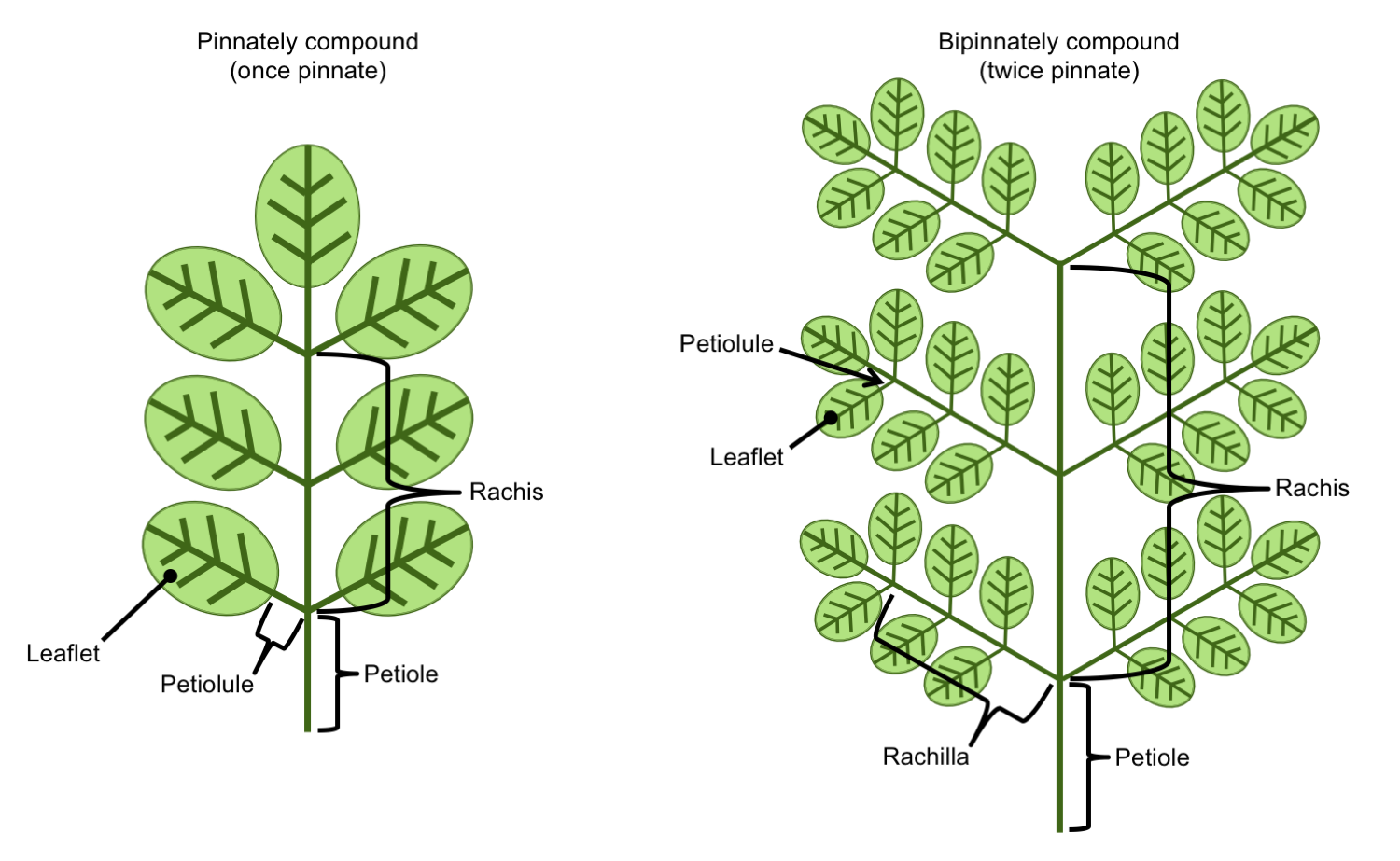
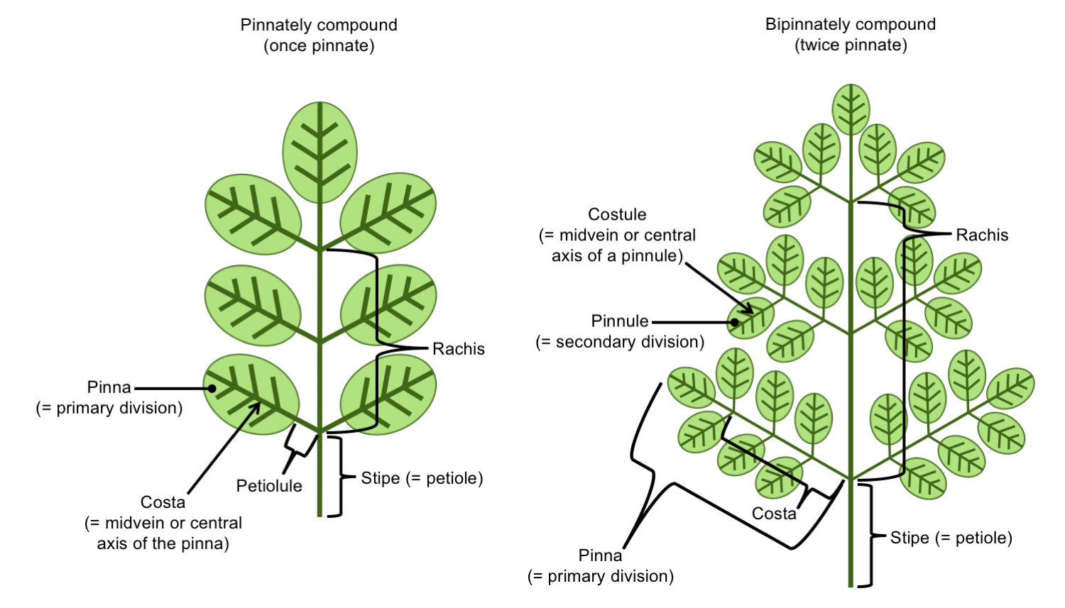
Overview By the end of this section, you will be able to do the following: Identify the parts of a typical leaf Describe the internal structure and function of a leaf Compare and contrast simple leaves and compound leaves List and describe examples of modified leaves

Labeled Diagram Of A Leaf
GCSE WJEC Structure of plants - WJEC Leaf structure Plants adapt in order to efficiently collect raw materials required for photosynthesis. These raw materials must be transported through the.
Leaf Structure & Evolution Digital Atlas of Ancient Life
Leaf Anatomy (Structure) Leaves are complex organs consisting of several layers serving various essential functions. They are the site of photosynthesis in plants, producing food. For cellular functions like photosynthesis and respiration, leaves require several cells and tissues to work in coordination.
Leaf Structure & Evolution Digital Atlas of Ancient Life
A diagram showing a leaf at increasing magnifications. Magnification 1: The entire leaf Magnification 2: Mesophyll tissue within the leaf Magnification 3: A single mesophyll cell Magnification 4: A chloroplast within the mesophyll cell Magnification 5: Stacks of thylakoids—grana—and the stroma within a chloroplast

Leaf Labelled Stock Photo Download Image Now iStock
Like the stem, the leaf contains vascular bundles composed of xylem and phloem (Figure 3.4.2.6 − 7 3.4.2. 6 − 7 ). When a typical stem vascular bundle (which has xylem internal to the phloem) enters the leaf, xylem usually faces upwards, whereas phloem faces downwards. The conducting cells of the xylem (tracheids and vessel elements.

Draw a labelled diagram of the external structure of a leaf. Brainly.in
Plant Leaves and Leaf Anatomy. Leaves are the site of photosynthesis in plants. Plant leaves help to sustain life on earth as they generate food for both plant and animal life. The leaf is the site of photosynthesis in plants. Photosynthesis is the process of absorbing energy from sunlight and using it to produce food in the form of sugars.

Leaf Structure Labeled Best Science Images and diagrams Pinterest Leaf structure and
Key points The leaf is one of the most important organs of a plant. Leaves produce food for the plant through a process called photosynthesis. The leaves of different plants vary widely in size,.